Last Chance to Catch NYC's Holiday Notalgia Train
We met the voices of the NYC subway on our nostalgia ride this weekend!


The American Museum of Natural History is one of the city’s most beloved institutions, with more than 200 scientists working across many disciplines. We previously covered the many secrets of this immense museum and showed you the art studios which create the incredible installations and dioramas, but today we are pleased to show you behind the scenes in Paleontology Division and the Microscopy and Imaging Labs. We learned how CT scanning and 3-D images are used to study everything from fossils to meteorites. As luck would have it, we gained entrance into the unique turret office of the Chair and Macaulay Curator of the Division of Paleontology – who happens to be the Curator of the next opening exhibit at the Museum, Dinosaurs Among Us. Below are a few of the photos we took on our behind-the-scenes look inside The American Museum of Natural History.
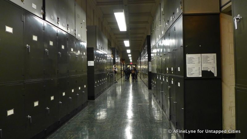
Back hallways within the Museum, one of the longest hallways in New York City, lined with lockers from floor to ceiling,
The Microscopy and Imaging Facility (featured below) is an interdepartmental lab which aids in the research of the Museum’s whole scientific community. The state-of-the-art imaging instruments include scanning electron microscopes (SEM), a transmission electron microscope (TEM), and an x-ray computed tomography scanner (CT Scanner). With this technology at hand, the lab has the ability to investigate internal features, using non-destructive methods. This allows for the exploration of priceless collections.
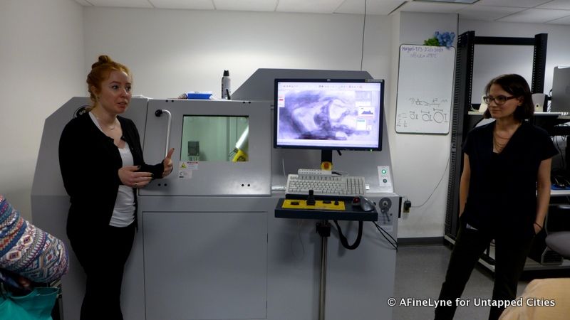
Amy Balanoff and Eugenia Gold, Research Associates at the Museum, standing aside a scan of a turkey fossil
The CT scanner (above) has the ability to capture thousands of sequential x-ray images, as if digitally dissecting. They have the ability to view high-resolution images of objects from fossils to meteorites. Most recently in the news was an amber fossil, donated to the Museum by a private collector, containing the world’s oldest known chameleon. With the use of the micro-CT scanner, scientists were able to view tiny bones and examine soft tissue – and confirm other features, such as the adhesive toe pads similar to those found in modern-day descendants. The discovery also suggests that the adaptation dates back more than 100 million years, which indicates that these creatures most likely didn’t get their start in Africa, as previously believed.
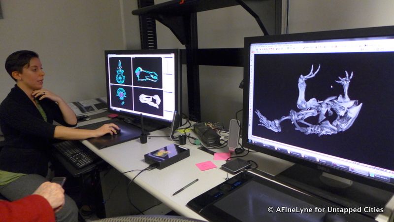
Exploring the evolution of the brain
In addition to a non-invasive view, the CT data can be used to reconstruct the central nervous system, and study the evolutionary history of the brain. 3-D representations of physical objects can be reconstructed, such as the interiors of fossilized skulls. These are known as digital endocranial casts, or endocasts.
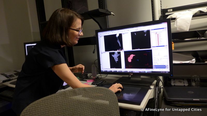
Viewing computer model of 3D reconstructions
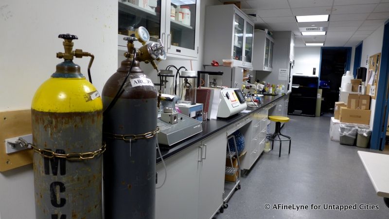
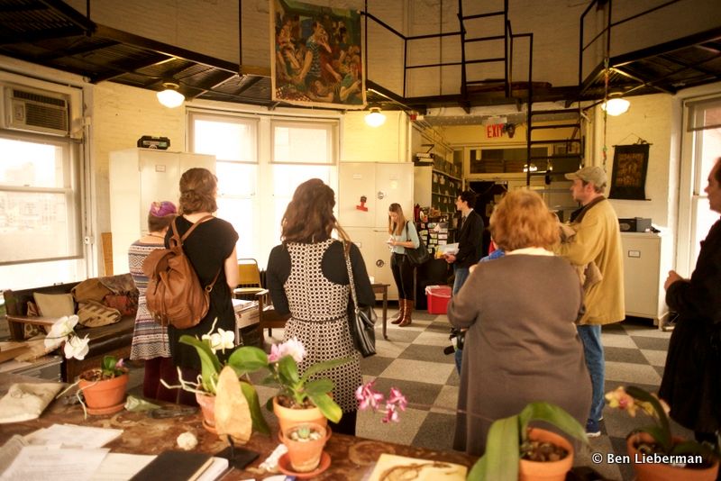
Entering the Microscopy and Imaging Facility at The American Museum of Natural History
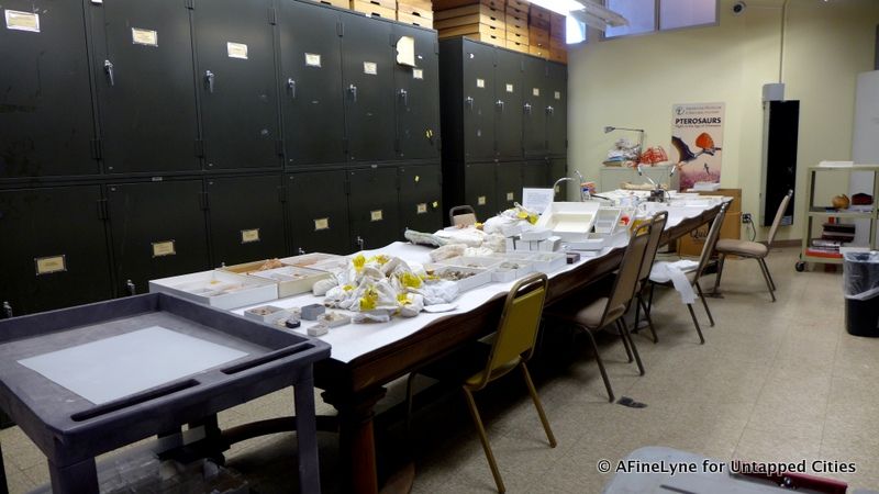
The Division of Paleontology – collection storage spaces
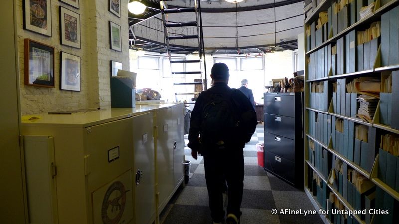
Entering the fourth-floor turret, office of Dr. Mark Norell
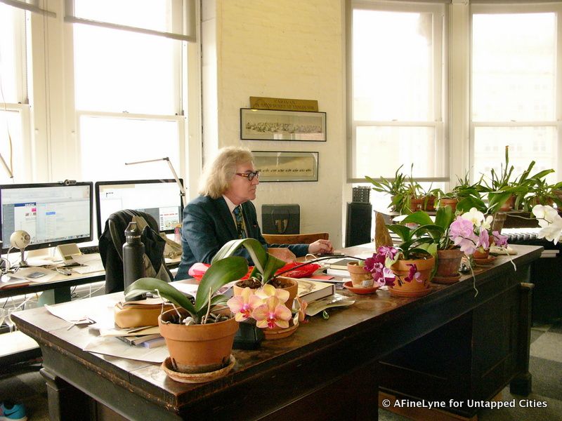
Entering a turret above the fourth-floor, on the north corner of the Museum on Central Park West, we found ourselves in the office of Dr. Mark Norell, Chair and Macaulay Curator, Division of Paleontology. The circular space, with soaring ceilings, has a spectacular view of Central Park from every window. File cabinets lined all the walls and orchids were blooming on his desk, window sills and side tables. Intermingled throughout the room were dinosaur specimens and Asian art.
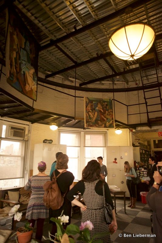
Dr. Norell’s love of art is reflected throughout his office
The view from behind Dr. Norell’s desk
Dr. Mark Norell, curator of Dinosaurs Among Us, has been one of the team leaders of the joint American Museum of Natural History/Mongolian Academy of Sciences expeditions to the Gobi Desert. With their discoveries, the team has generated new ideas about bird origins and the groups of dinosaurs to which modern birds are most closely related. He was part of the team who discovered Ukhaa Tolgod in 1993, and he was part of the team that in 1998 announced the discovery in northeastern China of two 120-million year old dinosaur species.
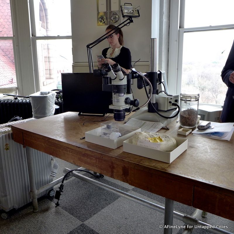
Microscope on a side-table in Dr. Norell’s office
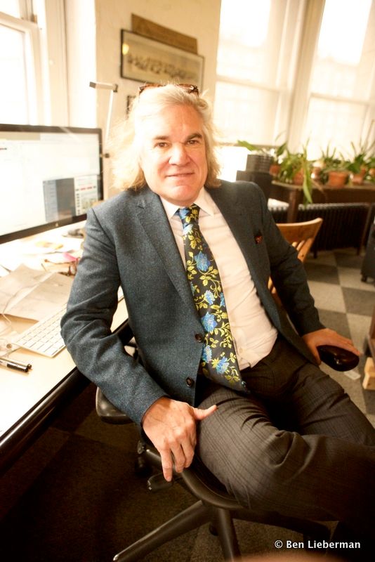
Dr. Mark Norell, Chair and Macauley Curator, Division of Paleontology, and Curator of Dinosaurs Among Us.
Continuing his work on evolutionary relationships of dinosaurs, Dr. Norell has developed new ways of looking at fossils using CT scans and imaging techniques, which we sampled in the Microscopy and Imaging Facility. His accomplishments too length to list, he continues to travel for new discoveries – last year to Asia seven times, Europe seven times, and commuting back and forth to China.
Get tickets to visit the American Museum of Natural History.
Read more about the Secrets of the American Museum of Natural History. Contact the author at AFineLyne.
Subscribe to our newsletter